ENDODONTIC RETREATMENT

28yr/ M with poorly obturated 36.

after Endodontic retreatment. 2 distal canals can be clearly seen.

16 yr. old female with large restoration over exposed pulp in 26. She has symptoms of apical periodontitis( tenderness to percussion and difficulty in biting). Periapical changes can be seen over disto-buccal root.

Biomechanical preparation is achieved with Protaper(rotary) and copious irrigation.
The tooth is obturated when it is asymptomatic.

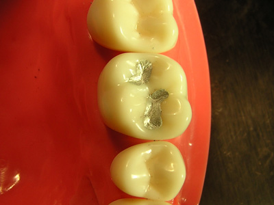
28 yr. old male with incomplete RCT in 36. The restoration sealing access cavity is broken. Remaining caries can also be seen.

After completion of endodontic treatment.

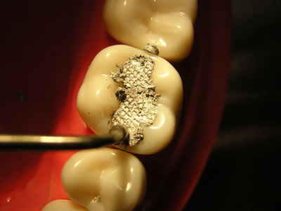
18 yr./F with amalgam restoration in 26. The restoration was done on exposed pulp chamber and the cement base can be seen extruding into it.

After completion of Endodontic treatment with Protaper(rotary).

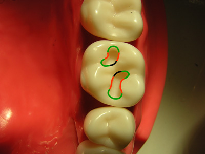
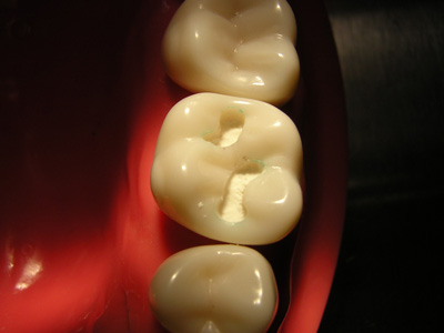
25 yr./F with carious exposure of 36.

After obturation.

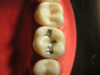
36 with PA radiolucency.

After Endodontic treatment. 2 distal canals can be seen.
PROCERA Bridge

30 yr. old female reports with existing bridge from 13 to 23. History reveals congenitally missing Upper lateral incisors. Metal Ceramic bridge was used to replace missing teeth. Patient has the following problems with existing bridge:

There is spacing distal to upper left canine which has not been addressed.
Additionally she had swelling in upper lips a month ago.
Patient requests replacement with Zirconia crowns (she read about them on the internet).

IOPA radiograph reveals a improper RCT on 11 and PA radiolucency on 21. A single GP point can be seen in 11. It also seems to be over-filed.

Removal of the bridge reveals grossly over-reduced teeth, particularly canines. The margins are not smooth.

Endodontics is completed on 11 & 21. The existing bridge is being used for temporization in between appointments.
After completion of RCT's, the margins are improved. Impressions are taken and sent to the lab. Zirconia cores are checked for fit and margins and returned to the lab.

The Procera bridges are back from the lab. They are intentionally constructed in two parts.

The white Zirconia cores can be seen.

A tooth has been added distal to 23. It serves only aesthetic purpose and is not in function.

The new bridge is cemented in place using RelyX Unicem. The gingival contours will gradually adapt to the new bridge. Problems of shape, size, colour and contour have been addressed. The incisal edges follow the lower lip contour(smile line). The teeth do not appear joined together.

Space closure has been achieved distal to 23.

The patient is visibly happy with her new bridge in place.
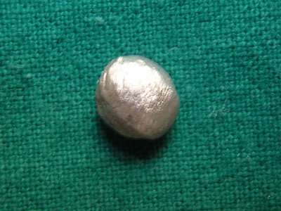
 Figure 1
 .
. Figure 3